Ebook Info
- Published: 2006
- Number of pages: 800 pages
- Format: PDF
- File Size: 97.34 MB
- Authors: Jack J. Kanski MD MS FRCS FRCOphth
Description
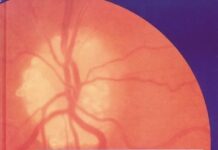
Highly Commended, RSM Awards 2007This book presents an encyclopedic visual catalogue of all clinical signs and symptoms in ophthalmology. It presents fully comprehensive coverage of all clinical conditions organised by region, starting with the front of the eye and working through to the retina in a very logical manner. The text is presented in a very readable and concise format, and is supplemented with over 2800 full colour clinical photographs of the highest quality. Virtually every condition (and variations within those conditions) has been illustrated. Clinical Diagnosis in Ophthalmology encourages the practitioner to view the patient as a whole to aid with the differential diagnosis of systemic and ocular signs. It comprehensively covers all systemic diseases associated with the eye. The illustrations are enhanced with brief, bullet-point style text that follows a tightly controlled, template format for quick and easy reference. Clinical Diagnosis in Ophthalmology provides an outstanding aid to clinical decision-making and practitioners will find the book very useful for matching conditions seen in the clinic. I will also be an ideal revision aid for examinations in ophthalmology. The book is also an extremely useful reference for all optometrists and primary care physicians.Serves as a complete diagnostic guide of ophthalmic disease.Features over 2,800 full-color clinical photographs, many original to Dr. Kanski’s private collection.Includes a CD-ROM containing all of the images from the text―suitable to download for electronic presentations.Provides concisely written, easy-to-read, templated chapters. Offers many angiograms, radiographs, and scans that emphasis pathological processes.Organizes topics logically―by anatomic region―starting with the front of the eye and progressing through to the retina, to make information easy to find.Represents the ideal guide for comparison to the full range of conditions seen in practice, or for certification/recertification review.
Reviews
Reviews from Amazon users which were colected at the time this book was published on the website:
⭐I am a 3rd year family medicine resident. I thought that as a brand new edition, this would be an update to the 2003 version,Clinical Opthalmology: A Systematic Approach also by Kanski. I was sorely mistaken. Much different than a textbook that gives you clinical information such has diagnosis and management that you can use, this book is almost 100% nothing but pictures, a glorified atlas. It is utterly useless for primary care practitioners. I would not be so disappointed in this had it not been so similarly titled as the previous, excellent text I mentioned. For all those who are not practicing ophthalmologists, the older text is definitely recommended above this.
⭐This is what an atlas ought to be. Pictures, pictures, and more pictures. This is not a textbook of ophthalmology but rather it is a picture atlas with multiple pictures of disease processes. The photos are of great quality and have an identifying caption. Thats it. Very little text.For those studying for your oral board certification exam, this is a great resource of photographs to use. The spectrum and volume of photos truly surpasses that found in the Spalton atlas as well as the Will’s atlas. Just remember, and I repeat myself, this is not a textbook.
⭐Simply one of the finest reviews of Ophthalmolgy in existense. This volume is packed with full color photos on every ophthalmic disease process imaginable as well as photos from the associated systemic diseases. There are external, slit lamp, fundus, periphery, IVFA, ICG, and radiagraphic photos for each diagnosis as indicated by clinical workup. Multiple photos of each disease demonstrate mild to severe manifestations, not just the classic worst case scenario. This book is designed primarily for Ophthalmologists, not primary care physicians. 90% of the diagnoses in this book require an Ophthalmologist’s consultation. The book is high quality and even includes a CD with all of the images available for use in presentations. My only gripe is that the limited text is written in the Queen’s English, so terms such as Oedma show up to irritate Americans. I do like the Spalton Atlas approach of pointing out the important findings in a line drawing next to the photo, and a slightly more in-depth discussion of the disease processes could be included, but the photo quality and quantity is simply unmatched. As the title implies, this is a clinical atlas- there are few or no microscopic pathology pictures. The stabismus section is also somewhat limited, but at less than $150 this atlas is a steal.
⭐I was a little disappointed as most of the photos showed advanced disease states. You would not likely see them in your office in Canada.
Free Download


